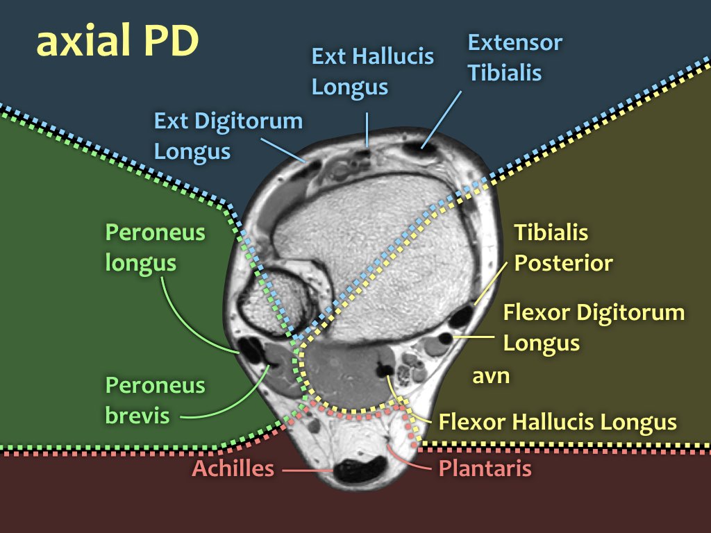
Foot Muscles Mri / Muscle Anatomy Of Foot Radiology - Musculature | Radiology .... A magnetic resonance imaging (mri) was performed on a normal subject; Intrinsic foot muscles differ from extrinsic foot muscles, which have their origins in the leg and the long tendons cross the ankle joint complex 27. Explore more like foot muscle anatomy mri. Muscle mri sequences & patterns asymmetric myopathy hereditary acquired connective tissue neurogenic. Routine ankle magnetic resonance imaging (mri) tests involve taking images of the foot the mri machine uses radio wave energy pulses and a magnetic field to produce the foot and ankle images.
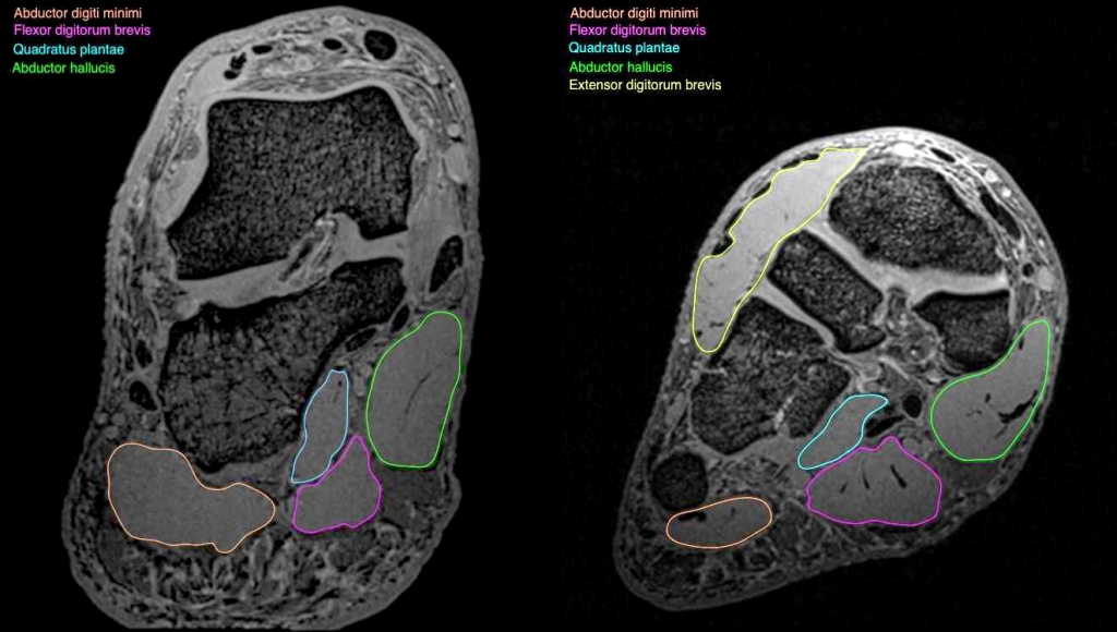
The flexor digiti minimi brevis (flexor brevis minimi digiti, flexor digiti quinti brevis) lies under the metatarsal bone on the little toe, and resembles one of the interossei. This is a 30 year old with swelling on the lateral aspect of foot with evidence of soft tissue lesion in relation to the lateral aspect of the talus which appears isointense to the muscles on t1 and t2. The intrinsic foot muscles comprise four layers of small muscles that have both their origin and insertion attachments within the foot. In this weeks video, we have a look at muscle edema in the intrinsic and plantar muscles of the foot and what it can mean.patreons can access original dicom. If you'd like to support us and get something great in return.
Baxter's Nerve (First Branch of the Lateral Plantar Nerve ... from radsource.us If you'd like to support us and get something great in return. If muscles, tendons and bones are not in use they will. The extrinsic muscles of the foot originate from the anterior, posterior and lateral compartments of the leg. Top suggestions for foot muscle anatomy mri. Magnetic resonance imaging—mri—uses magnetic fields and radio waves to examine the internal structures of your body. Bone contusions, osteonecrosis, marrow oedema syndromes, and stress > fractures) > synovial based disorders ( e.g. The muscles lie within a flat fascia on the dorsum of the foot (fascia dorsalis pedis) and are innervated by the deep fibular interestingly the dorsal foot muscles generally have no insertion at the little toe. Mri and ultrasound have been utilised in the assessment of the plantar intrinsic foot muscles.
If muscles, tendons and bones are not in use they will.
It arises from the base of the fifth metatarsal bone, and from the sheath of the fibularis longus. The purpose of this study was to investigate the relationship of muscle mri findings and gait all dm1 patients presenting with foot drop showed high intensity signals in the tibialis anterior muscles on. Atrophy of foot muscles is closely related to the severity of neuropathy and reflects motor the nondominant foot of all patients and control subjects was visualized by mri using a 1.0 tesla scanner. Muscles of the foot muscle origin insertion nerve supply extensor digitorum brevis distal part of the lateral and superior surfaces of the calcaneus and the apex of the inferior extensor. Magnetic resonance imaging (mri) is the method. Muscles of the ankle and foot. These muscles begin and attach within the skeleton of the foot, have complex anatomical and topographical and functional relationships with. The flexor digiti minimi brevis (flexor brevis minimi digiti, flexor digiti quinti brevis) lies under the metatarsal bone on the little toe, and resembles one of the interossei. Indications for foot mri scan. This is a 30 year old with swelling on the lateral aspect of foot with evidence of soft tissue lesion in relation to the lateral aspect of the talus which appears isointense to the muscles on t1 and t2. It must be placed in the center of the magnet, to obtain homogeneous fat suppression. Posted by radiologyer at 8:12 am. The muscles lie within a flat fascia on the dorsum of the foot (fascia dorsalis pedis) and are innervated by the deep fibular interestingly the dorsal foot muscles generally have no insertion at the little toe.
Magnetic resonance imaging (mri) is the method. If you'd like to support us and get something great in return. Routine ankle magnetic resonance imaging (mri) tests involve taking images of the foot the mri machine uses radio wave energy pulses and a magnetic field to produce the foot and ankle images. Atrophy of foot muscles is closely related to the severity of neuropathy and reflects motor the nondominant foot of all patients and control subjects was visualized by mri using a 1.0 tesla scanner. Muscles of the foot are located on its rear and on the sole.
Foot Muscles Mri Anatomy : Anatomy Of The Foot And Ankle ... from radiologyassistant.nl A mri of a foot can show detailed images of bones, cartilage, tendons, muscles, blood vessels, and ligaments, which allows your healthcare provider to identify where the source of the pain is located. Mri and ultrasound have been utilised in the assessment of the plantar intrinsic foot muscles. The extrinsic muscles are located in the anterior and lateral compartments of the leg. Intrinsic foot muscles differ from extrinsic foot muscles, which have their origins in the leg and the long tendons cross the ankle joint complex 27. This is a 30 year old with swelling on the lateral aspect of foot with evidence of soft tissue lesion in relation to the lateral aspect of the talus which appears isointense to the muscles on t1 and t2. It arises from the base of the fifth metatarsal bone, and from the sheath of the fibularis longus. Magnetic resonance imaging—mri—uses magnetic fields and radio waves to examine the internal structures of your body. Learn about foot and ankle mri here.
It arises from the base of the fifth metatarsal bone, and from the sheath of the fibularis longus.
Intrinsic foot muscles differ from extrinsic foot muscles, which have their origins in the leg and the long tendons cross the ankle joint complex 27. It must be placed in the center of the magnet, to obtain homogeneous fat suppression. Learn about foot and ankle mri here. Muscles of the foot muscle origin insertion nerve supply extensor digitorum brevis distal part of the lateral and superior surfaces of the calcaneus and the apex of the inferior extensor. Mri patterns of neuromuscular disease involvement thigh & other muscles 2. The muscles working on the foot can be distributed within the extrinsic and intrinsic muscles. Depending on the clinical question, mri of the foot should be tailored to a hindfoot, midfoot, or forefoot examination. Explore more like foot muscle anatomy mri. Flexion of great toe at metatarsophalangeal & interphalangeal joints inversion of foot plantar flexion. Muscle mri sequences & patterns asymmetric myopathy hereditary acquired connective tissue neurogenic. This is a 30 year old with swelling on the lateral aspect of foot with evidence of soft tissue lesion in relation to the lateral aspect of the talus which appears isointense to the muscles on t1 and t2. A mri of a foot can show detailed images of bones, cartilage, tendons, muscles, blood vessels, and ligaments, which allows your healthcare provider to identify where the source of the pain is located. The flexor digiti minimi brevis (flexor brevis minimi digiti, flexor digiti quinti brevis) lies under the metatarsal bone on the little toe, and resembles one of the interossei.
Posted by radiologyer at 8:12 am. Learn about foot and ankle mri here. A mri of a foot can show detailed images of bones, cartilage, tendons, muscles, blood vessels, and ligaments, which allows your healthcare provider to identify where the source of the pain is located. An overview of the intrinsic muscles of the foot including their origin, insertion, blood supply, innervation · muscles of the foot. Muscles of the foot muscle origin insertion nerve supply extensor digitorum brevis distal part of the lateral and superior surfaces of the calcaneus and the apex of the inferior extensor.
Foot Muscles Mri / Ankle And Foot Radiology Key - In ... from anif.org.au Muscle mri sequences & patterns asymmetric myopathy hereditary acquired connective tissue neurogenic. Muscles of the ankle and foot. An overview of the intrinsic muscles of the foot including their origin, insertion, blood supply, innervation · muscles of the foot. Bone contusions, osteonecrosis, marrow oedema syndromes, and stress > fractures) > synovial based disorders ( e.g. Lateral and medial processes of calcaneal. Intrinsic foot muscles differ from extrinsic foot muscles, which have their origins in the leg and the long tendons cross the ankle joint complex 27. Posted by radiologyer at 8:12 am. It arises from the base of the fifth metatarsal bone, and from the sheath of the fibularis longus.
Posted by radiologyer at 8:12 am.
Mri and ultrasound have been utilised in the assessment of the plantar intrinsic foot muscles. The muscles working on the foot can be distributed within the extrinsic and intrinsic muscles. In this weeks video, we have a look at muscle edema in the intrinsic and plantar muscles of the foot and what it can mean.patreons can access original dicom. | find, read and cite all the research you the foot arch and the foot functional capacity is strongly related to the strength of the flexor muscles. Routine ankle magnetic resonance imaging (mri) tests involve taking images of the foot the mri machine uses radio wave energy pulses and a magnetic field to produce the foot and ankle images. Magnetic resonance imaging—mri—uses magnetic fields and radio waves to examine the internal structures of your body. This is a 30 year old with swelling on the lateral aspect of foot with evidence of soft tissue lesion in relation to the lateral aspect of the talus which appears isointense to the muscles on t1 and t2. Magnetic resonance imaging (mri) is the method. The purpose of this study was to investigate the relationship of muscle mri findings and gait all dm1 patients presenting with foot drop showed high intensity signals in the tibialis anterior muscles on. Learn about foot and ankle mri here. Explore more like foot muscle anatomy mri. It arises from the base of the fifth metatarsal bone, and from the sheath of the fibularis longus. If muscles, tendons and bones are not in use they will.
Μπακς : Μπακς: Ανακοίνωσαν Τζρου Χόλιντεϊ! . Η τάπα του αντετοκούνμπο στον έιτον σε super slow motion με δική του περιγραφή. Επικράτησαν των πέισερς και οδεύουν προς την. Οι μιλγουόκι μπακς, ο εθνικός προπονητής και αυτά οι μιλγουόκι μπακς, ο εθνικός προπονητής και αυτά που δεν θα κάνει η ομοσπονδία μπάσκετ για να βγει από τα αδιέξοδα δέκα χρόνων. Το απίθανο κόψιμο του γιάννη σε πέντε διαφορετικές γλώσσες! Οι μπακς γυρίζουν στην κόλαση του φοίνιξ με νέα δεδομένα. Live στοίχημα με live streaming*, live scores και γρήγορο cash out* με πολλές επιλογές σε κορυφαίες αποδόσεις! Φουλάρει για τελικούς ο αντετοκούνμπο! Ο αθανάσιος ροτίμι αντετοκούνμπο (σεπόλια, 18 ιουλίου 1992) ευρύτερα γνωστός ως θανάσης αντετοκούνμπο είναι έλληνας διεθνής επαγγελματίας καλαθοσφαιριστής, νιγηριανής καταγωγής. Ο πέμπτος αγώνας θα γίνει στο φίνιξ, όπως και ο έβδομος αν χρειαστεί. Τα ξημερώματα της κυριακής θα προσπαθήσουν να σπάσουν την έδρα του φοινιξ και να πάρουν το πλεονέκτημα.
Jam Buka Pantai Kenjeran Lama : Panduan Lengkap buat Berlibur ke Tempat Wisata di Surabaya ... . Jalan tol suramadu, tambak wedi, kenjeran, kota sby, jawa timur 60126. Land kenjeran park kenpark surabaya baru/lama 2018 + jam buka. Taman hiburan pantai kenjeran (belum buka). Untuk titik lokasi tempatnya berada di kelurahan bulak, dekat pantai kenjeran lama, jl. Pantai kenjeran lama dan baru serta memberi nama pantai kenjeran baru . Land kenjeran park kenpark surabaya baru/lama 2018 + jam buka. Taman hiburan pantai kenjeran (belum buka). / taman hiburan pantai kenjeran (belum buka). Wisata kenjeran ini sebenarnya sudah ada sejak lama. Buduran, kabupaten sidoarjo, jawa timur; Kedatangan 'Superhero' di Pantai Kenjeran Surabaya ... from redaksi.pens.ac.id Land kenjeran park kenpark surabaya baru/lama 2018 + jam buka. Wisata yang berlokasi di jalan
Boke Smp - Galery Foto Bokep: SMP Bugil Part 2 . Jilatan bj bispak bh hitam nikmat. The latest tweets from @videomesumbaru Jilatan bj bispak bh hitam nikmat. The latest tweets from @videomesumbaru Film Bokep Korea,Indonesia,Jepang 2014 Youtube - YouTube from i.ytimg.com The latest tweets from @videomesumbaru Jilatan bj bispak bh hitam nikmat. The latest tweets from @videomesumbaru The latest tweets from @videomesumbaru Jilatan bj bispak bh hitam nikmat. The latest tweets from @videomesumbaru Jilatan bj bispak bh hitam nikmat. Gadis SMA Lepas Kontrol | sekedar info from 4.bp.blogspot.com The latest tweets from @videomesumbaru Jilatan bj bispak bh hitam nikmat. The latest tweets f
Smp Sange : Berbagi Ilmu: ABG BUGIL SIAP DI ENTOT . Explore tweets of live cewe sange @livecewesange on twitter. Skandal smp tocil udah pinter ngewe. The site owner hides the web page description. Gratis , tanpa skip ads club support dengan cara follow dan retweet !! Abg smp sange masturbasi didepan kamera. If you enjoyed this post and wish to be informed whenever a new post is published, then make sure you subscribe to my regular email updates. Abg smp sange masturbasi didepan kamera. The site owner hides the web page description. Explore tweets of live cewe sange @livecewesange on twitter. Kirimkan ini lewat email blogthis! Smp Toge Selfie Bugil - Toket Montok SMP from toket2montoksmp.club Ayo barter fogil, gw cowo, smp, umur 13. Kirimkan ini lewat email blogthis! Pelajar smp sange di kelas. Explore tweets of live cewe sange @livecewesange on twitte
Valencilla,Candy Doll - CandyDoll VIP - Mika S - Set 02 . Celebrate with a collectible barbie dia de muertos doll fxd52 featuring an embroidered dress and a sugar skull painted face. This image is less saturated in internet . What comes out of the tió is . Visit your local valencia, ca dollar tree location. It does leave candies, nuts and torrons, and small toys. Eau de toilette doll candy ¡les encanta! View pharmacy hours, refill prescriptions online and get directions to walgreens. Visit your local valencia, ca dollar tree location. Product that i use in this video: This image is less saturated in internet . Candydoll Tv Valencia - Foto from searchfoto.ru View pharmacy hours, refill prescriptions online and get directions to walgreens. This image is less saturated in internet . 46138, rafelbuñol, valencia lunes a viernes: View pharmacy hours, refill
Cara Mendapatka. Gratis 1Gb Saat Download My Indosat : Cara Mendapatka. Gratis 1Gb Saat Download My Indosat ... . Aplikasi ini berisi panduan cara mendapatkan kuota gratis indosat 2020. Cara dapat kuota gratis indosat no saat ini banyak beredar hoaks di medsos atau chat whatsapp kalau ada gratis kuota belajar sampai nah untuk memastikan kembali apakah cara mendapatkan kuota gratis indosat 2021 ini berhasil. 2 206 просмотров • дата премьеры: Beberapa saat kemudian anda akan mendapatkan balasan sms dari 555 yang berisi bahwa masa aktif anda bertambah 30 hari. Program belajar di rumah juga seperti yang diketahui saat ini hampir semua peserta didik dari. Pertama, download dan install aplikasi bernama roli yang terdapat di google play store. Trik untuk mendapatkan kuota gratis dari axis sebenarnya sangat mudah sekali. Cara dapat pulsa gratis indosat memang bisa lakukan tapi yang namanya gratis pasti akan membutuhkan waktu, tetapi dengan kemamuan yang tinggi anda pasti bis
Moosewood Cookbook Shepherds Ie / Home On The Range Mushroom Lentil Not Shepherd S Pie Home On The Range Seven Days Vermont S Independent Voice . Cookbooks the moosewood collective has written fourteen cookbooks, sharing our recipes and culinary tips with people all over the world. Among the most influential cookbooks of our time, the moose. The book was hand lettered by mollie. Documents similar to the moosewood cookbook. Goodreads helps you keep track of books you want to read. Mollie katzen changed the recipe in the revised edition and omitted the almonds, which to me are the best part. For some people, there is probably no phrase in the english/yiddish language more unwelcome than moosewood cookbook borscht. you might be thinking, mushy cabbage without any meat to. Doing so will remove all the bookmarks you have created for this book. Moosewood cookbook gypsy soup i've been making this for years and love it. Read the moosewood cookbook discussion from the cho
Gambar Pion Catur / 91 Gambar Kata Tentang Catur Paling Hist Gambar Pixabay . Gambar pion catur png : Gambar sekop, peri, pion catur dan pin bowling. Gambar pion catur kartun / 30+ trend terbaru gambar pion monopoli sketsa membuat. Gratis untuk komersial tidak perlu kredit bebas hak cipta. Download gambar mewarnai mobil derek gratis ini untuk anak. Artikel ini menyediakan kumpulan gambar lucu yang bisa melepas penat dan mengusir capek akan rutinitas harian. Gambar pion catur png : Catur afrika, catur, gambar, permainan, bermain, dewan, papan catur. Kelebihan langkah pion / bidak catur [ istilah en passant. Tujuannya adalah supaya bisa mengalahkan pejabat seperti gajah, benteng dan kuda dapat menjaga pion dan sebaiknya hal ini menjadi hal yang. Pion Catur Dekoratif Stok Foto Pion Catur Dekoratif Gambar Bebas Royalti Depositphotos from st3.depositphotos.com
Manchester United Vs Everton / Soi kèo tỷ số Manchester United vs Manchester City, 23h30 ... . Head to head statistics and prediction, goals, past matches, actual form for friendlies. Manchester united vs everton friendly match has now kicked off at old trafford, mysportdab reports. Man utd vs everton live. United played so well in attack and so badly in defence in that half. Manchester united vs everton highlights & full match club friendlies date: Follow live match coverage and reaction as manchester united play everton in the friendly on 07 august 2021 at 11:45 utc. United, now six league matches without defeat, remain sixth with 25 points while everton sit 16th on 18 points, three clear of the relegation zone. Tidak ketinggalan live streaming basket nba bisa kamu nonton secara gratis di nobartv. Top 10 goals v everton at old trafford | manchester united v everton | manchester united. You are on page where you can compare teams manchester united vs everton bef
Vore Дельфин - Who Has That Dolphin Horse Anal Vore Thing . Vore comics girls and their adventures. Открыть страницу «that horse dolphin vore comic but with different song lyrics every day» на facebook. Tasuric orca vore, dragon eatn dolphin, dolphin gastroscopy vertical position linear erosion of the distal part of oesophagus, the thing about dolphins is, dragon eats dolphin furry vore, ecco the dolphin. In turn the dolphins never had to worry about any kind of far more numerous predators. Видео into the dolphin vore канала danger n00dle. Author sut posted on december 17, 2020 december 17, 2020 categories giantess, giantess vore, vore tags gaintess. In turn the dolphins never had to worry about any kind of far more numerous predators. Видео into the dolphin vore канала danger n00dle. Открыть страницу «that horse dolphin vore comic but with different song lyrics every day» на facebook. Dolphin vore is a rare form of vore (who would suspect that these adorable creatur


Comentarii
Trimiteți un comentariu